responsive
Anatomy eand biomechanics
Rotator cuff tendons work as an engine for the shoulder.
Each tendon has a corresponding muscle around the Scapula (shoulder blade), and they are divided into three parts according to their anatomical location.
Five tendons are divided into three categories. They constitute separate units that attach the bone to the muscle:
- Upper side tendon: Supraspinatus Tendon
- Front tendons: Teres major and teres minor
- Back side tendons: Subscapularis tendon
These tendons work together to ensure movement and control of the arm .The biomechanics of the tendons are complex as they move and rotate in all planes of motion.
Joint strain is caused by repetitive motion, combined with age-related wear. The tendons can then become inflammed, causing tendinitis, and the tendons can tear partially or completely.
Clinical diagnosis - Treatment can be decided on in relation to the levels of pain felt by the patient. Pain and difficulties in moving the joint are key elements in diagnosis, at all stages of the condition.
Symptomes
responsive
Diagnosis of rotator Cuff injuries
Le diagnostique est d’abord clinique réalisé par un examen et un interrogatoire spécifique qui permet de suspecter une lésion tendineuse et complété par des examens complémentaires adaptés : radiographie, échographie IRM Arthroscanner
Rotator Cuff injuries treatment
-
Medical treatment aims first at pain management for young patients in the early stages of this condition, and who suffer from serious torn ligaments.
Patients are asked to rest the shoulder and avoid making any painful, or aggravating movements.
Medical treatment often includes pain killers and anti-inflammatory drugs (contraindications are taken into account).
-
Rehabilitation aims to alleviate pain and reinforce the affected tendons. Stretching exercises that the patient can do by themselves are used to strengthen the injured tendons. Sometimes balneotherapeutics are prescribed.
-
Surgery becomes an option if well managed medical treatment is not enough, and if tendons are not too retracted.
Patient, une prise en charge axée sur
Traitement
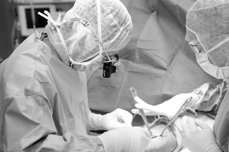
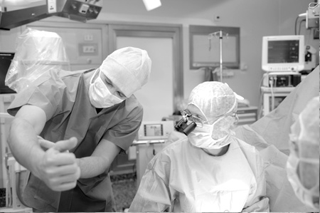
Tendon surgery techniques
Several techniques are used to fix tendons as close as possible to their original locations.
- A simple surgical debridement, or cleaning, of the tendons is carried out. This is done with the aid of cameras.
- Conventional, or open, surgery or by minimal invasive surgery.
- Arthroscopy
This technique is currently very popular because of high quality improvements in technology-assisted surgery.
This option presents several benefits: preservation of surrounding tissues, lower blood loss, better view of injuries and surrounding elements, lower risk of infections, absence of skin aggravation, and less painful and quicker post-surgery recovery.
After a preparation stage, tendons are fixed using anchors on the bone on which sutures are placed. They pass through holes in the anchor stabilizing the injury by this system of different sutures.
Other surgical procedures can also be carried out including bursectomy, or cleaning, of the subacromial space, additional removal or fixation of the intra-articular part of the tendon of the biceps, and acromioplasty, or removal of the distal inferior acromion process of the scapula. This can be performed if tendon impingement was detected in pre-op examinations.
Post surgery
- To avoid stiffness, post-op rehabilitation should begin early.
- Patients are hospitalized for 48 hours on average. Before surgery, an assessment by a rehabilitation centre might be done on a case by case basis.
- Out-patient treament, where the patient returns home the same day of surgery, is also possible in certain cases - as long as the same conditions as hospitalization are met.
- In all cases, pain management strategies are established after surgery according to the protocols written by our anaesthetists.
- During hospitalization post surgery visits, your surgeon will inform you about shoulder movement exercises.
Patient check-ups take place durint the 3rd week and 3rd and 6th month after surgery, in consultation with the surgeon. The check-up includes an x-ray of the tendon and a clinical self-assessment questionnaire.
Full functional recovery usually occurs 6 months after surgery, when the patient resumes work.
Video with ELKHOLTI MD and VOGELS MD.